Ludwig’s angina causes, symptoms, management
Contents
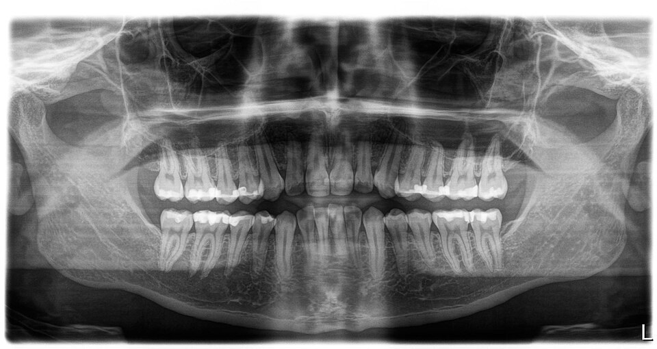
Photo by Farhang Amini, CC BY 4.0, via Wikimedia Commons
Ludwig's angina is a serious bacterial infection of the floor of the mouth, under the tongue. It is a rapidly spreading cellulitis that affects the spaces on both sides of the floor of the mouth and under the jaw: the submandibular, sublingual and submental spaces.
Despite the name, it is not related to heart angina. It most often follows an infected tooth, especially a lower second or third molar, or an injury to the mouth. It is uncommon in children.
This condition is treatable with prompt care. Without treatment, swelling can block the airway, which is life-threatening. It needs emergency treatment to protect the airway and careful monitoring.
Symptoms of Ludwig's angina
This infection often follows a tooth infection, mouth injury or other infection in the mouth. The area can swell quickly. Symptoms of Ludwig's angina include:
- Neck pain
- Drooling of saliva
- Difficulty swallowing
- Pain or tenderness in the floor of the mouth, below your tongue
- A muffled "hot potato" voice or trouble speaking
- Fatigue or confusion
- Swelling of the neck
- An earache
- Redness of the neck
- Tooth pain
- Chills
- Swelling of the tongue, which can push the tongue up and back
- Fever
As the infection progresses, it can cause trouble breathing. It may also lead to life-threatening complications such as sepsis.
If you have trouble breathing, trouble swallowing saliva or rapidly growing swelling under your jaw, call 911 or go to the emergency room right away.
Is Ludwig's Angina Contagious?
Ludwig's angina itself is not spread from person to person. It develops when bacteria already in your mouth, usually from an infected lower molar, spread into the deep tissues.
Once the infection develops, it can spread quickly between the spaces of the floor of the mouth and neck. In severe cases, it can travel along the deep tissue layers of the neck into the chest.
That spread can cause serious complications such as mediastinitis (infection in the middle of the chest), jugular vein thrombosis, fluid around the lungs or heart, necrotizing fasciitis and sepsis.
Causes of Ludwig's Angina
The most common cause of Ludwig's angina is a dental infection. The infection is usually caused by a mix of mouth bacteria, including streptococci, staphylococci and anaerobic bacteria. Causes and risk factors include:
- Poor dental hygiene
- Mandible fracture
- Tooth abscess
- Trauma or lacerations in the mouth
- Infections of lower second and third molar
- A recent tooth extraction
- Submandibular gland infection
Penetrating injury of the floor of the mouth
HOW IS LUDWIG'S ANGINA DIAGNOSED? A doctor examines the head and neck for swelling and redness. Tests may include:
1- Dental X-ray
Infections at the roots of the teeth, such as a tooth abscess, can be identified with a dental X-ray.
2- CT scan
A CT scan of the neck with contrast is used to see how far the infection has spread. If spread into the chest is suspected, a chest scan is done too.
3- Cell Culture
A sample of fluid or pus from the infected area may be tested to identify the bacteria.
Differential Diagnosis
Some conditions look similar and must be ruled out. These include a hematoma under the tongue, angioedema (sudden allergic swelling) and cancer of the tongue.
Treatment of Ludwig's Angina
The first priority is keeping the airway open. This may mean placing a breathing tube, often with a flexible scope while the patient is awake, or in some cases an emergency tracheostomy.
1- Systemic
Antibiotics are given, usually through a vein at first and then sometimes by mouth. The infected tooth also needs dental treatment.
2- Incision and drainage
Surgery may be needed to drain fluid or pus, especially if the infection doesn't improve with antibiotics. Drainage can be done:
- Intraoral (through the mouth)
- Externally (through the skin of the neck)
External drainage is used if the infection spreads to the submandibular space.
Intraoral drainage may be enough if the infection is still limited to the sublingual space. In external drainage, a surgeon makes an incision under the jaw and opens the deep tissue spaces with a blunt hemostat. Very often, the surgeon finds swollen, fluid-filled tissue rather than frank pus.
3- Tracheostomy
A tracheostomy, a surgical opening in the windpipe, is done when the airway is in danger of being blocked.
With prompt airway care, antibiotics and surgery when needed, most people recover. A 2023 review notes that death rates fell from over 50% before antibiotics to about 8% with modern treatment.
HP Thoughts: Many surgical blotches occur because of so many things, one of which is vanity. Do you really need to undergo dental reconstruction? Read our next post and tell us what you think: Smile Makeover.
Sources
- Ludwig angina, MedlinePlus
- Ludwig's Angina, Tunis Med (PMC)